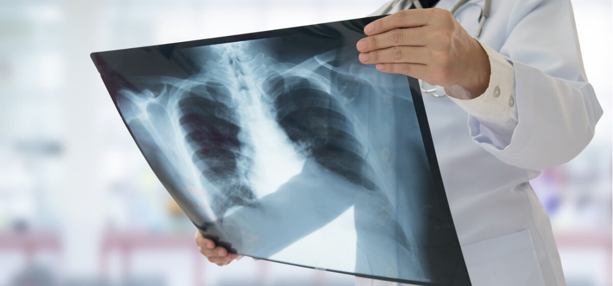
What Do My Chest X-Ray Results Show?

A chest X-ray is a procedure that produces images of the inside of the chest. It helps doctors assess the condition of the lungs, heart and chest wall. The process of producing a chest X-ray is fast and easy, so it’s especially useful in evaluating injuries in emergency situations. It’s also one of the first procedures performed to evaluate symptoms such as chest pain, a persistent cough or difficulty breathing.
What are Chest X-rays Used to Diagnose?
There are many reasons for a chest X-ray to be ordered, including chest X-ray heart. A chest X-ray may be prescribed to investigate a number of different symptoms, including:
- Chest pain
- Fever
- Persistent cough
- Shortness of breath
Doctors use chest X-rays to provide information that helps with the diagnosis or assessment of a variety of conditions. Wondering what chest X-rays can detect or asking, “What does a chest X-ray show?” The procedure can reveal:
- The condition of the heart. Changes or abnormalities in the size or shape of the heart can indicate heart valve problems, heart failure or fluid around the heart.
- The health of the lungs. Chest X-rays can reveal or provide details on a collapsed lung, pneumonia and other infections, cancer, emphysema, cystic fibrosis and other conditions that affect the lungs.
- The condition of certain blood vessels in the chest. The outlines of large blood vessels like the aorta, pulmonary veins and arteries can be seen in a chest X-ray. Providers use the X-ray to detect issues like aneurysms or other problems.
- Bone fractures. Fractures to the spine or ribs, and other problems with those bones, can be viewed with a chest X-ray.
- Positioning of a pacemaker or other device. Chest X-rays allow doctors to assess the position and functioning of pacemakers and defibrillators (devices that keep the heart rhythm normal) as well as catheters (tubes used to deliver medication or for dialysis).
- Calcium deposits. The presence of calcium in the heart or blood vessels can point to damaged heart muscle or valves and the associated increased risk of a heart attack. In the lungs, calcium is often an indicator of past infections.
How is a Chest X-ray Performed?
A chest X-ray is a quick and painless procedure. In most cases, the patient stands against a recording plate and a technician takes images of the chest from two perspectives: one from the back and another from the side. Chest X-rays are ready for review by a doctor almost immediately.
Benefits and Risks of Chest X-Rays
X-rays provide many benefits. This includes providing doctors with rapid access to helpful diagnostic information and providing patients with a simple, pain-free procedure. However, it’s understandable to ask, “How much radiation in a chest X-ray?” The answer is that the amount of radiation is small and that X-rays are very safe. However, if you’re pregnant or believe you might be pregnant, it’s important to let your care team know before a chest X-ray is performed.
Chest X-Ray Results
For people wondering, “Will a chest X-ray show lung cancer?” it’s helpful to know that following a chest X-ray, a specially trained physician called a radiologist evaluates the images from the procedure and produces a report about the chest X-ray findings. The referring doctor uses this report to prescribe treatment in the case of abnormal chest X-ray images. Or, when there are normal chest X-ray findings, and the chest X-ray results interpretation reaches the conclusion “chest X-ray normal,” the doctor shares that no treatment is needed.
Baptist Health provides safe and efficient inpatient and outpatient diagnostic imaging for adults and for children (imaging for children available at only some locations). As one of the area’s most advanced diagnostic imaging centers, our services are fully integrated with our excellent medical care. In this way, we work together to detect the earliest signs of disease or injury and provide expert treatment.