Types of Diagnostic Medical Imaging Explained
Medical imaging is the process of creating visual representations of internal organs, bones, abnormal growths or inflammations, and other structures or areas. It is used for many purposes including for assessing the functioning of the body’s systems and developing a diagnosis in the case of injury or disease.
Types of Diagnostic Medical Imaging Explained
There are many forms of medical imaging, each using a different type of technology or technique to achieve a particular result. Some of the more common types of medical imaging are:
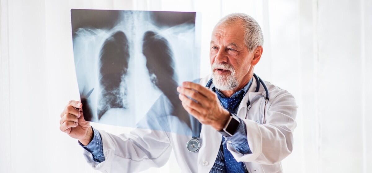
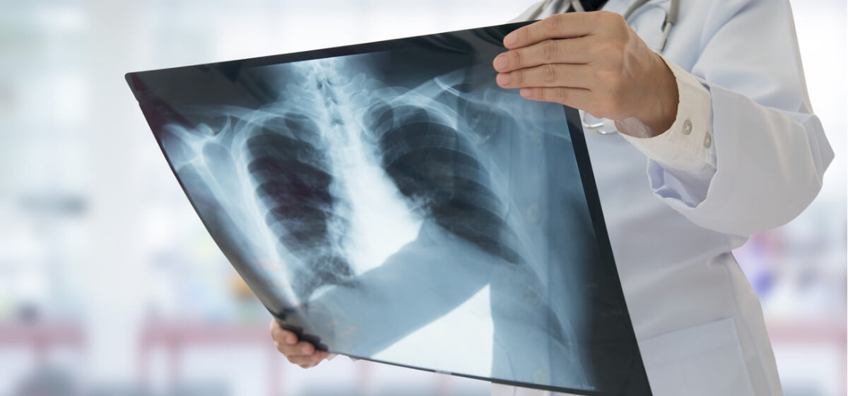
X-ray radiography
Electromagnetic radiation, in the form of X-rays, is projected toward a part of the body and captured behind the body on film or by a detector. The difference in how much energy is absorbed by body structures produces a two-dimensional image. People are most familiar with X-rays in the context of assessing damage to bones.
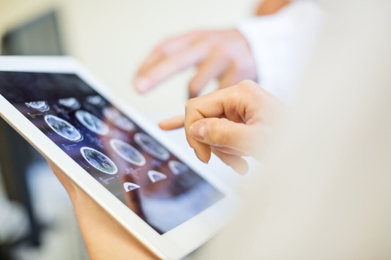
Computed tomography (CT) Scan
Multiple X-ray images produced from different angles are processed by a computer to produce a 3D representation of the area or structure. CT scans are often used in emergency medicine to determine if a patient has internal injuries or bleeding.
Ultrasound
Sound waves, with frequencies higher than humans can hear, are focused on an area of the body. The waves are reflected in varying degrees by varying densities of tissues and used to create an image that can be viewed on a monitor. Many people are familiar with ultrasound from its use with pregnant mothers. It is also used to examine muscles, tendons, blood vessels, and internal organs.
Magnetic resonance imaging (MRI)
Powerful magnetic fields and radio waves, rather than radiation, are used to create an image of bones, organs, blood vessels, and other soft tissues. MRIs are often used to assess conditions in the chest, abdomen, and pelvis.
Endoscopy
A camera is inserted into the hollow area within an organ or a body cavity and captures photos and/or video of the area. A colonoscopy is one common use of endoscopy.
Thermography
Infrared waves are captured by a special camera to assess blood flow or inflammation in an area of the body. This type of imaging is frequently used in the diagnosis of sports injuries.
Positron emission tomography (PET)
Typically used to detect cancer, PET introduces a small amount of radioactive sugar (called a tracer) into the body. Cancer cells need more energy than normal cells and digest sugars for that energy. As the cancer cells metabolize the sugars, the PET camera detects the metabolic activity to create a visualization of internal structures.
Why Medical Imaging is Important
Medical imaging is critical to the care and treatment of patients for a number of reasons. It is a non-invasive way to evaluate the condition or functioning of an organ, system or area of the body. Consequently, it eliminates the pain and discomfort that would be caused by invasive procedures.
Medical imaging also enables healthcare teams to detect and assess injuries or disease at early stages, making treatments more effective. It is faster and more efficient than other forms of assessment, which helps control healthcare costs.